
✨ Advances in AI Imaging Algorithms for Early Lung Cancer Screening
🩺 The Core Role of AI Imaging in Early Lung Cancer Screening
Computer-Aided Detection (CADe) and Diagnosis (CADx) systems integrated with Deep Learning (DL) have demonstrated significant clinical value in early lung cancer screening. By enhancing the detection rate of early pulmonary oncology signs—such as Ground-Glass Opacities (GGOs)—these systems facilitate the clinical translation of diagnostic findings.
In terms of workflow optimization, AI systems act as a “second reader” for radiologists. Through pre-screening and preliminary image analysis, AI effectively alleviates the diagnostic workload, potentially shortens turnaround times (TAT), and thereby elevates overall operational efficiency.
📌 Technical Mechanisms of Deep Learning in Micro-Nodule Identification
Deep learning algorithms based on Convolutional Neural Networks (CNNs), trained on large-scale CT imaging datasets, are capable of learning and capturing complex texture features and spatial distribution patterns that are subtle to the human eye. These algorithms hold a distinct advantage in identifying micro-nodules smaller than 5 mm in diameter, helping to mitigate false-negative rates caused by visual fatigue or lesion size.
Clinical validation data indicates that AI-assisted interpretation can boost radiologists’ sensitivity to pulmonary nodules by approximately 8% to 15%, highlighting the immense potential of AI in improving early lesion detection.
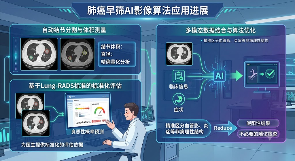
🧬 Clinical Applications and Accuracy Optimization
In clinical practice, AI systems automate nodule segmentation and volumetric measurement while predicting malignancy probabilities based on the Lung-RADS framework, offering physicians standardized evaluation criteria.
Cutting-edge algorithms are also incorporating multi-modal data—combining patient medical histories and clinical records—to differentiate non-pathological structures such as vascular shadows or inflammation. This approach actively reduces false-positive rates and avoids unnecessary follow-up examinations.
💊 Demystifying Common Misconceptions About AI in Lung Cancer Diagnosis
- Misconception 1: AI can completely replace physicians.At present, AI functions strictly as an assistive tool designed to support physicians in image analysis and clinical decision-making. Ultimate diagnostic authority and therapeutic decision-making must remain with certified radiologists and clinicians. AI cannot replicate the role of a physician in managing complex cases, patient-doctor communication, or humanistic care.
- Misconception 2: AI screening offers absolute accuracy.AI systems are not infallible and are subject to false-negative or false-positive outcomes. System performance can be influenced by multiple variables, including image quality, cross-vendor CT hardware variances, and training dataset limitations. Consequently, AI screening results cannot serve as an absolute diagnostic gold standard and must be interpreted alongside comprehensive clinical assessments.
🔬 Current Limitations and Knowledge Gaps
Despite its promising potential in early screening, AI deployment currently faces several key hurdles:
- Long-Term Prognostic Evidence: There is a lack of robust prospective studies directly proving that AI-assisted screening significantly improves long-term prognostic indicators, such as the five-year survival rate.
- Algorithm Generalization Challenges: Due to inherent image data discrepancies generated by CT scanners from different manufacturers (e.g., GE, Siemens, Philips), the cross-device generalization capability of AI algorithms requires further validation.
- Ethical and Economic Considerations: The legal frameworks defining liability for AI diagnostic misses remain ambiguous. Furthermore, cost-effectiveness analysis (CEA) data regarding the large-scale deployment of AI systems is still insufficient, warranting further evaluation of its economic viability.
✨ Evidence-Based Medicine (EBM) Backing
- The Lancet Digital Health has published meta-analyses regarding the diagnostic accuracy of AI.
- Radiology has featured relevant studies on the application of deep learning in Low-Dose Computed Tomography (LDCT) screenings.
- Official reports and white papers from the Radiological Society of North America (RSNA) and the American College of Radiology (ACR) provide established guidelines and expert consensus on the clinical application of AI in thoracic imaging.
⚠️ Disclaimer: This content is provided for informational and educational purposes only and does not constitute medical advice or diagnostic criteria. Please consult a qualified healthcare professional for medical consultation.
© Satoko International Medical Support Co., Ltd. All Rights Reserved. | Unauthorized reproduction is strictly prohibited.